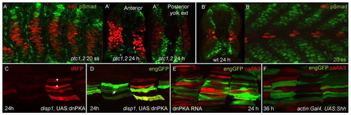
Modulation of Smad accumulation and Eng expression by Hh and BMP pathway activity. (A-A′) Parasagittal optical section and transverse sections at different levels along the rostrocaudal axis of ptc1hu1602;ptc2tj222 double homozygous mutant embryo at 20 ss and 24 hours, respectively, showing vastly expanded MPs (revealed by 4D9, red) and reciprocal loss of pSmad accumulation (green) compared with wild-type embryo at a similar stage (B,B′). Note expansion in domain of pSmad accumulation dorsally and ventrally in ptc mutants, especially at more caudal levels; compare with 24 hour wild-type embryo (B′). (C,D) Parasagittal optical sections of Tg(actin:GAL4);Tg(eng2a:eGFP)i233; disp1tf18bmutant embryo injected with UAS:dnPKA;tRFP at one- to two-cell stage. Most tRFP expressing fibres are also eGFP+ve, consistent with an autonomous effect of Hh pathway activity on eng2a induction. Note that not all fibres expressing tRFP (C) ectopically activated eng2a:eGFP (*); conversely, one eGFP+ve fibre (marked by an arrow in D) was tRFP–ve; this possibly represents a fibre induced by residual Hh signalling activity, as is sometimes observed in disp1tf18bmutant embryos. (E,F) Merged images of 24 hours Tg(eng2a:eGFP)i233 embryos expressing caALK3 under actin:GAL4 control in which the Hh pathway has been activated by injection of dnPKA mRNA or of UAS:Shh DNA. (E) dnPKA injection results in the ectopic activation of the eng2a:eGFP transgene (green) but this is suppressed in cells expressing caALK3 (red). Similarly, (F) late expression of Shh activates the eng2a:eGFP transgene in fast twitch fibres but this is suppressed by caAlk3 (red). On average, only 1.4 out of 13 tRFP+ve fibres also expressed eGFP (n=22 hemisegments from seven embryos).
|
