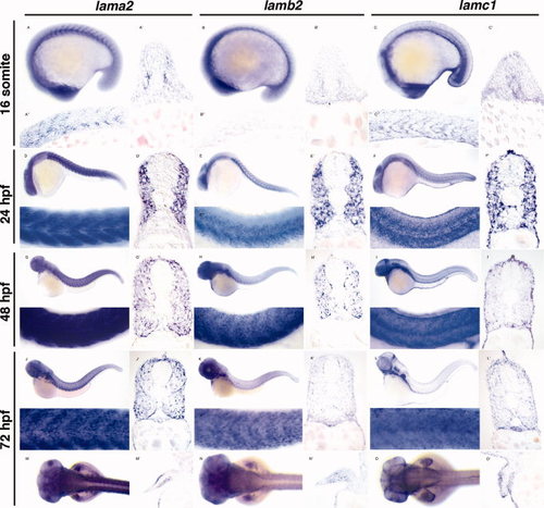
Expression of the major muscle laminin genes (lama2, lamb2, and lamc1) in zebrafish during development. A–C: Lateral whole-mount view of the 16–somite stage showing lama2 (A), lamb2 (B), and lamc1 (C) expression, with corresponding transverse (A′–C′) and sagittal (A″–C″) sections in the developing myotome. D–F′: Lateral whole-mount view of larvae staged 24 hr post-fertilization (hpf) is shown for lama2 (D, D′), lamb2 (E, E′), and lamc1 (F, F′) with corresponding transverse (D′–F′) sections through the trunk musculature. All genes are expressed in the slow and fast compartments of the myotome. G–I″: Lateral view of larvae staged at 48 hpf is shown for lama2 (G, G″), lamb2 (H, H″), and lamc1 (I, I″) with corresponding transverse (G′–I′) sections through the trunk musculature. All genes are expressed in the slow and fast compartments of the myotome with additional expression in neuronal tissues. J–O′: Larvae staged at 72 hpf are shown in lateral (lama2 [J, J″], lamb2 [K, K″], and lamc1 [L, L″]) and dorsal (lama2 [M], lamb2 [N], and lamc1 [O]) view with corresponding transverse sections through the trunk (J′–L′) and fin (M′–O′) musculature. All genes are expressed in the slow and fast compartments of the myotome and in the pectoral fins. In addition, lama2 is expressed in the posterior hypaxial muscle covering the yolk (M).
|
