
Fig. 1
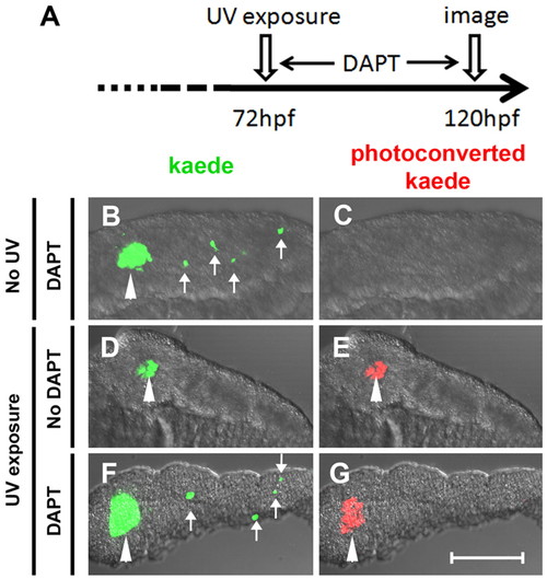
β-Cells of the secondary islets do not originate from principal islet β-cells. (A) Schematic of experimental timeline. The green fluorescent kaede protein in β-cells of ins:kaede fish was photoconverted to red fluorescent protein by UV exposure at 72 hpf. (B-G) Rendered confocal images of microdissected pancreata from ins:kaede fish at 120 hpf. (B,F) DAPT treatment from 72-120 hpf induces secondary islets (white arrows), including differentiated β-cells that are marked by kaede protein. (E,G) Photoconversion at 72 hpf labeled all β-cells present at that time point with red fluorescence. By 120 hpf, photoconverted kaede is still apparent in principal islet cells (white arrowheads) but is not detected in DAPT-induced secondary islets (G). Scale bar: 100 μm. |