
FIGURE
Fig. 6
- ID
- ZDB-FIG-110121-34
- Publication
- Yu et al., 2011 - The Cell Adhesion-associated Protein Git2 Regulates Morphogenetic Movements during Zebrafish Embryonic Development
- Other Figures
- All Figure Page
- Back to All Figure Page
Fig. 6
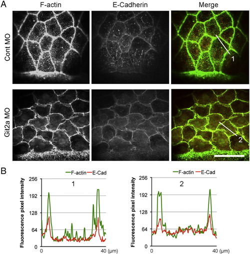
E-Cadherin localizes to adherens junctions in git2a morphants. (A) Representative immunofluoresence images of cortical actin (green) and E-Cadherin (red) in EVL cells of control and git2a morphant embryos. Scale bar, 50 μm. (B) F-actin and E-Cadherin fluorescence pixel intensity profiles of control (1) and Git2a morphant (2) embryos. |
Expression Data
Expression Detail
Antibody Labeling
Phenotype Data
| Fish: | |
|---|---|
| Knockdown Reagent: | |
| Observed In: | |
| Stage: | 75%-epiboly |
Phenotype Detail
Acknowledgments
This image is the copyrighted work of the attributed author or publisher, and
ZFIN has permission only to display this image to its users.
Additional permissions should be obtained from the applicable author or publisher of the image.
Reprinted from Developmental Biology, 349(2), Yu, J.A., Foley, F.C., Amack, J.D., and Turner, C.E., The Cell Adhesion-associated Protein Git2 Regulates Morphogenetic Movements during Zebrafish Embryonic Development, 225-237, Copyright (2011) with permission from Elsevier. Full text @ Dev. Biol.