
Fig. S2
- ID
- ZDB-FIG-101208-18
- Publication
- Gorsi et al., 2010 - Dynamic expression patterns of 6-O endosulfatases during zebrafish development suggest a subfunctionalisation event for sulf2
- Other Figures
- All Figure Page
- Back to All Figure Page
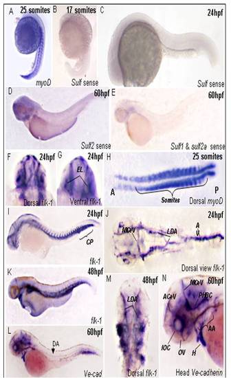
Expression of sulf sense probes and endothelial and muscle markers during zebrafish development. Representative images of sulf sense probes at 17 somite stage (B), 24hpf (C)and 48hpf (D,E). Markers used as positive control for all WISH experiments, myoD expression at 17 somite stage (A) dorsal ciew of somite staining(H). Expression pan-endothelial marker flk-1 at 24hpf lateral view of whole embryos (I) and dorsal view of flat mount of (J), dorsal (F) and ventral view (G) of head at 24hpf. Lateral view of flk-1 expression at 48hpf (K) and dorsal view (M). Lateral view of embryo expressing ve-cad at 60hpf (L) and close up of the head of embryo (N). AA, aortic arches; ACeV, anterior cerebral vein; AV, axial vessels; CP; caudal plexus; E, eye; EL, eye lens; H, heart; IOC, inner optic circle; LDA, lateral dorsal aortae; MCeV middle cerebral vein; OV, optic vein; PHBC, primordial hindbrain channel; PICA, primitive internal cartoid artery; PrA, prosencephalic artery. |