Fig. 5
- ID
- ZDB-FIG-101123-8
- Publication
- Joseph, 2004 - Zebrafish IRX1b in the embryonic cardiac ventricle
- Other Figures
- All Figure Page
- Back to All Figure Page
|
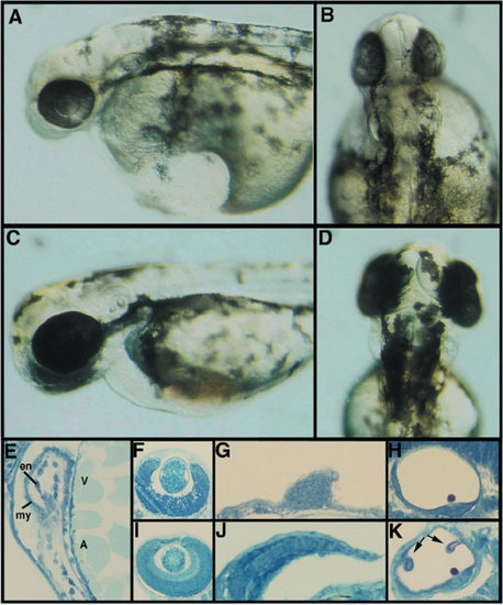
Phenotype of embryos injected with a morpholino designed to block IRX1b translation. A: Injection of an IRX1b morpholino (IRX1b-MO) antisense oligonucleotide prevents proper central nervous system development as shown here in these lateral views of a live 2 days postfertilization (dpf) embryo injected with a high dose of 1,000 μM. B: Dorsal view of the same IRX1b morpholino-injected embryo. C: A wild-type sibling of the morpholino-injected embryo shown in A and B. D: Dorsal view of the same wild-type embryo. E: IRX1b-MO–injected embryos develop with a distinctive ventricular (V) and atrial cardiac chamber (A); these chambers both have an endocardium (en) and a myocardium (my). F: Methylene blue azure II stained sections of IRX1b morpholino-injected embryos show other embryonic structures to be disrupted. The eye of IRX1b-MO–injected embryos has an altered retinal laminar pattern (compare with I). G: Fin outgrowth is reduced (compare with J). H: Semicircular canals do not form in the otic vesicles of IRX1b-MO–injected embryos, but otoliths do form (compare with K). I: Section through the eye of a wild-type sibling shows the normal laminar pattern of retinal neurons. J: Section through a wild-type fin shows the extent of fin outgrowth as well as the usual cell arrangement within the fin. K: Section through a wild-type sibling otic vesicle shows the presence of semicircular canal precursors (arrows). |
| Fish: | |
|---|---|
| Knockdown Reagent: | |
| Observed In: | |
| Stage: | Long-pec |

