
Fig. 1
- ID
- ZDB-FIG-101122-58
- Publication
- Ozbudak et al., 2010 - Spatiotemporal compartmentalization of key physiological processes during muscle precursor differentiation
- Other Figures
- All Figure Page
- Back to All Figure Page
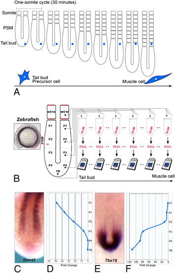
Generation of a microarray time series covering early stages of muscle differentiation in the zebrafish embryo. (A) A descendent of the tail-bud precursor cells (blue cell) enters the PSM posteriorly and progressively matures as it become located more and more anteriorly in the PSM during axis elongation. The cell eventually becomes incorporated into a somite, where most cells differentiate into muscles in zebrafish embryos. (B) Generation of the microarray time series. (Left) Lateral view of a 12-somite zebrafish embryo showing the position of the PSM (pink box). The samples used for the microarray time series were collected from six consecutive spatial positions in the PSM and recently formed somites on both sides of an embryo. Microarray expression profiles (D and F) faithfully recapitulate the expression of Dmrt2 (C) and Tbx16 (E) detected by in situ expression in 15-somite zebrafish embryos. Expression levels (D and F) were normalized by the expression levels in the tail bud (TB) and the somites (S1), respectively, to show the fold increase or decrease, respectively, along the spatial axis. |