|
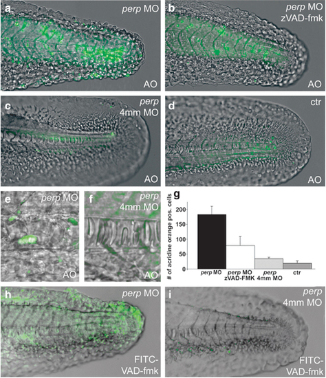
Loss of Perp causes caspase-dependent apoptosis. Acridine orange (a–f) and FITC-VAD-fmk (h,i) stainings of live embryos at 30 hpf; (a,c,e,h) injected with perp MO, (b) injected with perp MO and treated with caspase inhibitor zVAD-fmk, (c,f,i) injected with perp 4 mm MO, (d) uninjected control; (a–d,h,i) lateral view on tip of tail; (e,f) magnified lateral view on notochord. Note that in the notochord of perp morphants (e), all cells display altered, roundish shapes, while only some of the cells have become acridine orange positive. (g) Diagram illustrating average numbers of acridine orange-positive cells in embryos as shown in (a–d) within the tail region posterior of the anus. Number of evaluated embryos: control n=5; perp 4 mm MO-injected n=5; perp MO-injected n=6; perp 4 mm MO-injected+zVAD-FMK n=8
|