|
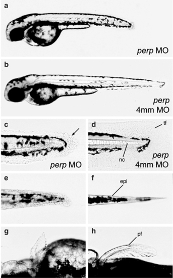
Loss of Perp causes tissue degeneration. All images show live embryos at 48 hpf, either injected with a perp-specific antisense MO (perp MO) (a,c,e,g), or the four mismatch control MO (4 mm) (b,d,f,h). (a,b) Overviews over entire embryos. (c–f) Higher magnifications of the tail region, (c,d) lateral views, (e,f) ventral views. The morphology of cells of the median fin fold and the notochord are changed in morphants (c; arrow points to roundly shaped cell), as compared to control embryos (d). The same is true for the trunk skin (e,f). (g,h) Dorsal view on the pectoral fins. Abbreviations: epi, skin; pf, pectoral fin; tf, tail fin
|