
Fig. 3
- ID
- ZDB-FIG-101119-16
- Publication
- Dahl Ejby Jensen et al., 2009 - Nitric oxide permits hypoxia-induced lymphatic perfusion by controlling arterial-lymphatic conduits in zebrafish and glass catfish
- Other Figures
- All Figure Page
- Back to All Figure Page
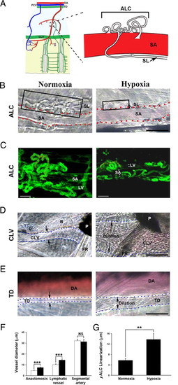
Hypoxia-induced linearization of arterial-lymphatic conduits (ALCs), lymphatic dilation, and blood perfusion in zebrafish and K. bicirrhis. (A) Schematic presentation of the ALC in the adult K. bicirrhis, with an expanded view of an ALC and its associated segmental artery (SA). DA, dorsal aorta; CLV, collecting lymphatic vessel; PCV, posterior cardinal vein; SL, segmental lymphatic; TD, thoracic duct. (B) Bright-field micrographs of ALCs in K. bicirrhis under normoxia and hypoxia. Black boxes show the ALCs. Dashed lines mark the SAs. (Scale bar, 50 μm.) (C) ALCs are found in the zebrafish tail region close to the SA and the lymphatic vessel (LV), as shown by EGFP-positive structures in fli1:EGFP zebrafish. ALCs budded from SAs and appeared as “tangled” compact corkscrew vascular plexuses (red dashed lines). Under hypoxic conditions, the compact, tangled architecture became linearized. (Scale bar, 20 μm.) (D) Hypoxia-induced dilation and blood perfusion in the CLV. Dashed blue lines mark the border of the CLV. B, bone; P, pigment; FR, fin ray. (Scale bar, 100 μm.) (E) Dilation of the TD under hypoxic conditions. Dashed blue lines mark the border of the TD, which became perfused under hypoxic conditions. (F) Quantification of averages of vessel diameters of the ALC, lymphatic vessel, and segmental artery under normoxia and hypoxia. ***, P < 0.001. NS, not significant. The open bars presents the values under normoxia and filled bars indicate the values under hypoxia. (G) Quantification of linearization of ALCs under normoxia and hypoxia. **, P < 0.01. |