
Fig. S7
- ID
- ZDB-FIG-101117-8
- Publication
- Padmanabhan et al., 2009 - Cardiac and vascular functions of the zebrafish orthologues of the type I neurofibromatosis gene NFI
- Other Figures
- All Figure Page
- Back to All Figure Page
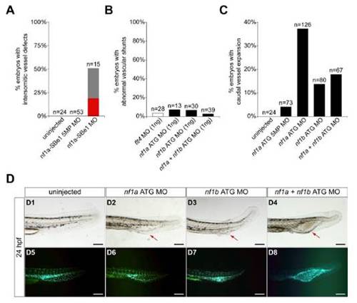
MO knockdown of nf1a, nf1b, or both together result in vascular defects at 24 at 48 hpf. (A) Quantification of vasculature at 24 hpf in uninjected and morphant Tg(fli:negfp)y7 (endothelial-specific nuclear GFP transgenic) zebrafish embryos. Uninjected, nf1a-SBe1 5MP (1 ng), and nf1a-SBe1 (1 ng) morphant embryos were qualitatively assessed for presence, absence (red), or an intermediate phenotype (gray) with regard to the developing trunk intersomitic vessels between somites 17–30 at 24 hpf. (B) Quantitation of vascular shunt phenotype seen with low (1 ng) doses of flt4, nf1a, nf1b, or nf1a + nf1b MOs. (C) Quantitation of cystic expansion phenotype observed in morphant Tg(fli:negfp)y7 embryos treated with 2 ng of the indicated MO(s). (D1–D8) Treatment of Tg(fli:negfp)y7 zebrafish embryos with an nf1a ATG MO, nf1b ATG MO, or nf1a + nf1b ATG MOs leads to a cystic expansion in the region of the caudal artery and vein (arrows, D2–D4) not present in uninjected embryos (D1). The expanded tissue was confirmed to be vascular by GFP expression (D6–D8). (Scale bars: 50 μm.) |