|
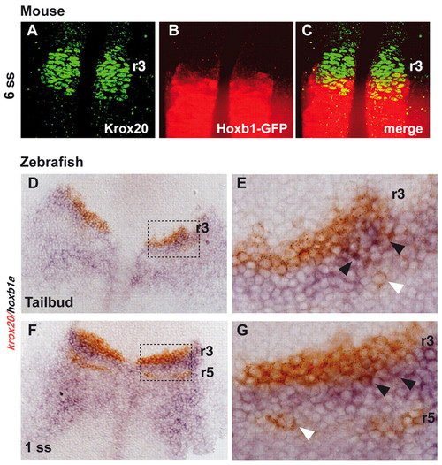
Overlap between Krox20 and Hoxb1 expression domains at the r3-r4 border. (A-C) Optical sections through the hindbrain of a whole-mounted Hoxb1GFP/+ mouse embryo at the 6-somite stage, immunolabelled for Krox20 (green) and Hoxb1-GFP (red): (A) anti-Krox20 immunofluorescence, (B) anti-GFP immunofluorescence and (C) merge. (D-G) Dorsal views of flat-mounted zebrafish embryos at the tail bud (D,E) or 1-somite (F,G) stages, hybridized with krox20 (red) and hoxb1a (blue). Black arrowheads point to double-labelled cells at the r3-r4 border. White arrowheads point to cells expressing krox20 at the level of prospective r5. E and G are higher magnifications of the outlined regions in D and F, respectively.
|