|
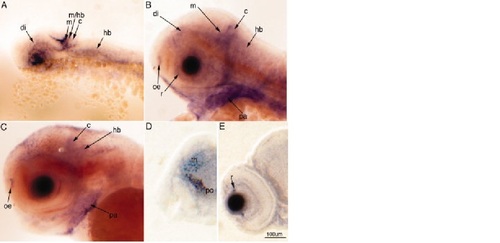
Cabin1 is expressed in the developing zebrafish nervous system. The developmental Cabin1 gene expression pattern as determined by mRNA in situ hybridization (ISH) in whole embryos is shown. A–C: Lateral views of the head at 24–72 hours postfertilization (hpf). D,E: Coronal sections through stained embryos at 72 hpf. A: At 24 hpf, expression is observed in the diencephalon (di), midbrain (m), midbrain–hindbrain boundary (m/hb), cerebellum (c), and hindbrain (hb). B: At 48 hpf, Cabin1 expression expands to the ganglion cell layer of the retina (r), and the olfactory epithelium (oe), and is also prominently expressed extraneuronally in the pharyngeal arches (pa). C,D: By 72 hpf, expression within the nervous system is still observed in the olfactory epithelium, cerebellum, and hindbrain, as well as in the diencephalon (C; in D, thalamus [th], preoptic area [po]). E: Expression in the retina at 72 hpf, has diminished to regions that correspond to regions where new neurons are generated.
|