
Fig. 5
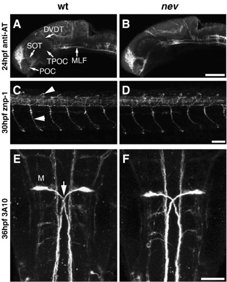
Early axon pathfinding appears normal in nev. Confocal projections in WT (A, C, E) and nev (B, D, F) stained with anti-acetylated tubulin (A and B), znp-1 (B and E), and 3A10 (C and F). Lateral views (A–D) and dorsal views (E and F). Anti-acetylated tubulin labels early axon pathways at 24hpf including the anterior commissure (AC), post-optic commissure (POC), supra-optic tract (SOT), tract of the post-optic commissure (TPOC), dorso-ventral diencephalic tract (DVDT), and medial longitudinal fasciculus (MLF). No differences are detectable between WT (A) and nev (B). Axon trajectories of primary motor neurons at 30hpf are normal in nev (D) compared to WT (C), including middle primary motor neurons (MiPs, upper arrowhead) and caudal primary motor neurons (CaPs, lower arrowhead). (E and F) Mauthner neurons and their axons (arrow in E) project normally in nev (F) as compared to WT (E) at 36hpf. Scale bar in B = 100 μm, D, F = 50 μm. |
| Antibodies: | |
|---|---|
| Fish: | |
| Anatomical Terms: | |
| Stage Range: | Prim-5 to Prim-25 |
Reprinted from Developmental Biology, 344(2), Pittman, A.J., Gaynes, J.A., and Chien, C.B., nev (cyfip2) Is required for retinal lamination and axon guidance in the zebrafish retinotectal system, 784-794, Copyright (2010) with permission from Elsevier. Full text @ Dev. Biol.