
Fig. 3
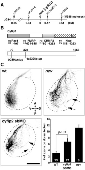
nevermind encodes Cyfip2. (A) Meiotic map of nev/cyfip2. Using a combination of SSLPs and SNPs, nev was fine-mapped to a 0.51 cM interval on LG14. Numbers above show recombinations in 588 meioses; numbers below indicate distance in cM. (B) Structure of Cyfip2. Domains known to be important for protein interactions with Cyfip1 and Cyfip2 are shown (see text for references). cDNA sequencing from tr230b and ta229f mutant embryos identified premature stop codons 79 amino acids (tr230b) and 328 amino acids (ta229f) into the protein. (C) A cyfip2 splice-blocking morpholino phenocopies nev. Dorsal views showing dorsonasal axons projecting onto the optic tectum. Dashed lines indicate approximate outline of the tectum. Dorsonasal axons in nev project inappropriately onto the dorsal half of the optic tectum, as do dorsonasal axons in embryos injected with a splice-blocking morpholino (SBMO) to cyfip2 (arrows). To quantify these errors, the number of axons projecting through the dorsal optic tectum was counted in wt, cyfip2 SBMO, and nev at 3dpf. Numbers of embryos shown inside bars. Scale bar = 50 μm. |
| Fish: | |
|---|---|
| Knockdown Reagent: | |
| Observed In: | |
| Stage: | Protruding-mouth |
Reprinted from Developmental Biology, 344(2), Pittman, A.J., Gaynes, J.A., and Chien, C.B., nev (cyfip2) Is required for retinal lamination and axon guidance in the zebrafish retinotectal system, 784-794, Copyright (2010) with permission from Elsevier. Full text @ Dev. Biol.