
Fig. 1
- ID
- ZDB-FIG-100706-29
- Publication
- Stephens et al., 2010 - Loss of adenomatous polyposis coli (apc) results in an expanded ciliary marginal zone in the zebrafish eye
- Other Figures
- All Figure Page
- Back to All Figure Page
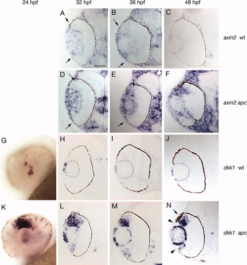
Expression of Wnt/β-catenin pathway genes in wild-type and apc mutant eyes. A,B: In 32 and 36 hours postfertilization (hpf) wild-type (wt) eyes, axin2 is expressed in the lens and in peripheral cells of the eye (arrows indicate central border of expression). C: By 48 hpf, only a few most peripherally located cells express axin2. D,E: In 32 and 36 hpf apc mutant eyes, axin2 is up-regulated in cells that express axin2 in wt eyes. F: In 48 hpf apc mutant eyes, axin2 expression is expanded, as it fails to become restricted to only a few peripheral cells. Even in apc mutants axin2 is never activated in the central retina. G-J: in 24-48 hpf wt eyes, only cells in the anterior lens epithelium express the Wnt inhibitor and Wnt target dkk1. K: In contrast, in 24 hpf apc mutant eyes dkk1 is strongly up-regulated in dorsal peripheral regions of the retina. L-N: Beginning at 32 hpf, expression is also observed in the ventral retina. N: Between 36 and 48 hpf dkk1 is down-regulated in the most peripheral cells (arrowheads). Throughout all stages observed dkk1 is also up-regulated in the lens. The dorsal retinal pigmented epithelium is expanded adjacent to the Wnt/β-catenin activation domain (arrow). |