
- Title
-
Loss of adenomatous polyposis coli (apc) results in an expanded ciliary marginal zone in the zebrafish eye
- Authors
- Stephens, W.Z., Senecal, M., Nguyen, M., and Piotrowski, T.
- Source
- Full text @ Dev. Dyn.
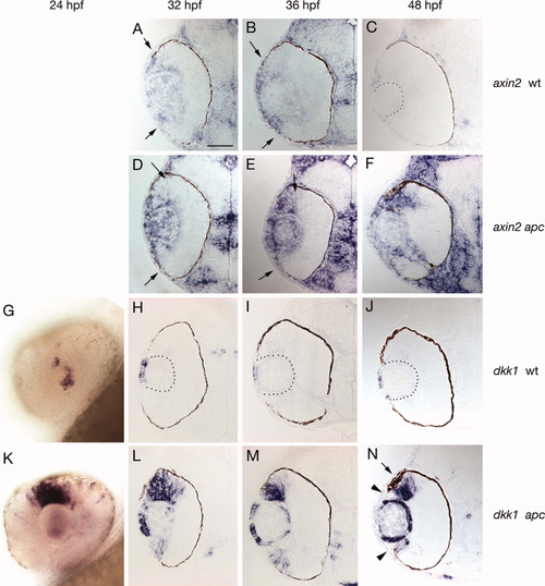
Expression of Wnt/β-catenin pathway genes in wild-type and apc mutant eyes. A,B: In 32 and 36 hours postfertilization (hpf) wild-type (wt) eyes, axin2 is expressed in the lens and in peripheral cells of the eye (arrows indicate central border of expression). C: By 48 hpf, only a few most peripherally located cells express axin2. D,E: In 32 and 36 hpf apc mutant eyes, axin2 is up-regulated in cells that express axin2 in wt eyes. F: In 48 hpf apc mutant eyes, axin2 expression is expanded, as it fails to become restricted to only a few peripheral cells. Even in apc mutants axin2 is never activated in the central retina. G-J: in 24-48 hpf wt eyes, only cells in the anterior lens epithelium express the Wnt inhibitor and Wnt target dkk1. K: In contrast, in 24 hpf apc mutant eyes dkk1 is strongly up-regulated in dorsal peripheral regions of the retina. L-N: Beginning at 32 hpf, expression is also observed in the ventral retina. N: Between 36 and 48 hpf dkk1 is down-regulated in the most peripheral cells (arrowheads). Throughout all stages observed dkk1 is also up-regulated in the lens. The dorsal retinal pigmented epithelium is expanded adjacent to the Wnt/β-catenin activation domain (arrow). |
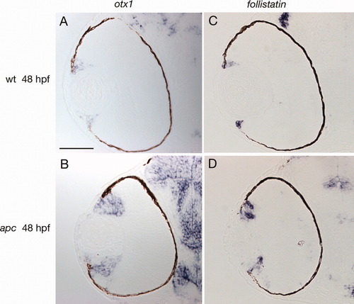
Peripheral fate markers are expanded in apc mutant eyes. A: otx1 is only expressed in the most peripheral cells in wild-type 48 hours postfertilization (hpf) eyes. B: in 48 hpf apc mutant eyes otx1 is centrally expanded. C: follistatin labels even fewer cells in the periphery of 48 hpf wild-type eyes than otx1. D: Like otx1, follistatin is expanded in 48 hpf apc mutant eyes. |

Wnt/β-catenin signaling inhibits retinal identity and proneural gene expression. A: The neural competence factor sox2 is expressed adjacent to the domain of Wnt/β-pathway activation and is absent from the most peripheral cells (arrows). B: The sox2 domain is centrally shifted in 48 hours postfertilization (hpf) apc mutant eyes. The more peripheral, expanded zone of Wnt/β-pathway activation is devoid of sox2 expression. C: The proneural gene atoh7 is expressed in a wedge adjacent, and more central to the sox2 domain. D: In 48 hpf apc mutant eyes, the atoh7 domain is centrally displaced. E,G: Double fluorescent in situ analysis of sox2 in red and atoh7 in green in 38 and 48 hpf wild-type eyes reveals that sox2 and atoh7 are expressed in adjacent, mutually exclusive domains. F,H: In 38 hpf apc eyes, the sox2 and atoh7 expression domains are shifted toward the central retina. In 48 hpf apc eyes, sox2 and atoh7 expression becomes more disorganized. I,J: Immunofluorescence with Zn8 shows that retinal ganglion cells differentiate in 48 hpf mutant eyes, even though they are restricted to the central retina. |

Wnt/β-catenin causes up-regulation of genes that inhibit neural differentiation. A: The transcriptional repressor her6 is expressed in the most peripheral cells in 48 hours postfertilization (hpf) wild-type eyes, as well as in cells surrounding the optic stalk. B: In apc mutant eyes, the her6 expression domain is expanded. C,D: her6 is likely a target of stat3 that is also up-regulated in apc mutant eyes. |
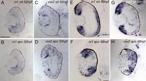
Wnt/β-catenin signaling is not involved in the initiation of vsx2 and rx1 expression but affects their maintenance. A,B: At 32 hours postfertilization (hpf), rx1 is broadly expressed in wild-type and apc mutant eyes, even though Wnt/β-catenin target genes are already expanded at this stage. C,D: In 32 hpf wild-type eyes, vsx2 is more strongly expressed in all cells of the dorsal, whereas in apc mutant eyes, it is also present in the ventral domain (D). E: In 48 hpf wild-type eyes, rx1 is expressed in all peripheral cells, encompassing the otx1, sox2, and atoh7 domain, as well as differentiating photoreceptor cells. G: In 48 hpf wild-type eyes, vsx2 is expressed in an overlapping domain with the exception of photoreceptor cells. F,H: In 48 hpf apc mutant cells, rx1 and vsx2 are expressed in more central cells but are down-regulated in the expanded CMZ. |

Proliferation is reduced in cells with active Wnt/β-catenin signaling. A: In 48 hours postfertilization (hpf) wild-type eyes, cyclinD1 is expressed in the sox2 and atoh7 domain but is excluded from the most peripheral cells. B: myca is expressed in the sox2 domain and is also excluded from the most peripheral cells. C,D: In 48 hpf apc eyes, cyclinD1 and myca are excluded from the expanded Wnt/β-catenin activation domain but are expressed in the central retina. E: In 50 hpf bromodeoxyuridine (BrdU) -treated wild-type embryos chased for 1 hr, BrdU is incorporated in cells of the sox2, atoh7, and cyclinD domain. Cells peripheral to these zones are mostly devoid of BrdU labeled cells (arrow heads). F: In 50 hpf apc mutant eyes, the expanded peripheral zone is BrdU-negative suggest that cells in the Wnt/β-catenin pathway domain are postmitotic or slowly cycling (outlined by arrowheads). G: In 50 hpf BrdU treated wild-type embryos chased for 2 hr, most cells in the periphery of the eye incorporated BrdU, suggesting that they are more slowly cycling than central cells. However, a few very peripheral cells are still BrdU negative. The very bright cells in the periphery are macrophages and not part of the developing retina. H: In 50 hpf apc mutant eyes that have been treated with BrdU and chased for 2 hr, BrdU is incorporated in single cells in 35% of all sections counted (n = 31; white arrowhead); demonstrating that not all cells are postmitotic. |

A-D: meis1 and pax6a act upstream of Wnt/β-catenin pathway activation, as these genes are normally expressed in the periphery of 48 hours postfertilization (hpf) apc mutant eyes. |

Cell death is not responsible for gene expression changes in apc mutant eyes. A,B: TUNEL (terminal deoxynucleotidyl transferase-mediated deoxyuridinetriphosphate nick end-labeling) staining of 48 hours postfertilization (hpf) wild-type (A) and apc mutant (B) eyes. In apc mutant eyes, more cells are undergoing cell death than in wild-type eyes. C,D: In situ hybridization of 48 hpf p53 morpholino injected wild-type and apc mutant embryos with the Wnt/β-catenin pathway target lef1. D: Even in the absence of cell death, apc mutant eyes show an expansion of Wnt/β-catenin target gene expression. |