
Fig. 4
- ID
- ZDB-FIG-100628-49
- Publication
- Cerveny et al., 2010 - The zebrafish flotte lotte mutant reveals that the local retinal environment promotes the differentiation of proliferating precursors emerging from their stem cell niche
- Other Figures
- All Figure Page
- Back to All Figure Page
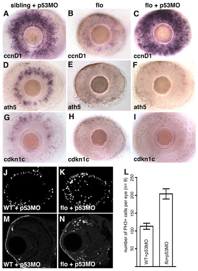
flo CMZ cells fail to transition from proliferation to differentiation, even in the absence of apoptosis. (A-I) Intact retinae from 3-dpf wild-type (A,D,G) and flo (B,E,H) zebrafish embryos and flo embryos injected with p53MO (C,F,I), labelled to show expression of the genes indicated bottom left. flo CMZ cells robustly express the proliferative marker ccnd1 (C) but not markers of cell cycle exit (F,I) when p53-mediated apoptosis is inhibited. (J,K,M,N) Confocal projections of laterally viewed wild-type + p53MO (J) and flo + p53MO (K) whole retinae and coronal sections of wild-type + p53MO (M) and flo + p53MO (N) retinae, all stained with anti-phosphohistone H3 (PH3). flo + p53MO eyes exhibit increased proliferation in/near the CMZ. (L) flo + p53MO embryos contain significantly more mitotic cells than wild-type + p53MO siblings (P<0.0001, Student′s t-test). The average number of mitotic retinal cells per eye (n=9 eyes from 9 different fish, examples shown in J,K) was calculated and graphed with error bars (95% confidence limits). |