
Fig. 4
- ID
- ZDB-FIG-100525-5
- Publication
- Sarmah et al., 2010 - Sec24D-dependent transport of extracellular matrix proteins is required for zebrafish skeletal morphogenesis
- Other Figures
- All Figure Page
- Back to All Figure Page
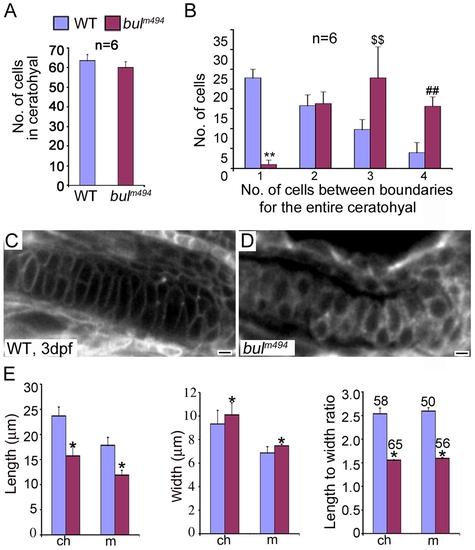
Analysis of chondrocyte shape and numbers in bulldog mutants. (A) The number of cells in the ceratohyals at 5 dpf is not significantly different between wild-type and bulldog embryos (counted in single optical plane of Alcian blue stained preparations, six different animals each). (B) In contrast, the number of cells spanning the entire width of the ceratohyal at 5 dpf is notably higher in bulldog. (C,D) Single-pass confocal images of the Meckel′s cartilage in live embryos marked with membrane tethered GFP tracer. Bulldog mutants (D) show multiple stacked chondrocytes as compared to a single spanning cell in wild-types (C). (E) The average chondrocyte width is comparable between wild-type and bulldog, whereas the length and the length-to-width ratio are significantly lower in the mutants. Cellular dimensions were counted in Meckel′s (m) and ceratohyal (ch) cartilages in three different live embryos at 3 dpf. The number of cells used for measurements is indicated in the right graph (E). * denotes p<0.0001; **, p<0.0001; $$, p<0.003; ##, p<0.0001. |
| Fish: | |
|---|---|
| Observed In: | |
| Stage Range: | Protruding-mouth to Day 5 |