Fig. S3
- ID
- ZDB-FIG-100525-12
- Publication
- Sarmah et al., 2010 - Sec24D-dependent transport of extracellular matrix proteins is required for zebrafish skeletal morphogenesis
- Other Figures
- All Figure Page
- Back to All Figure Page
|
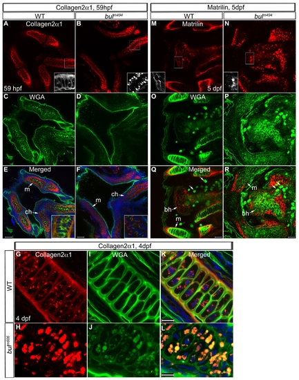
Cartilage Matrix Proteins are not Trafficked in bulldog Chondrocytes. Loss of Sec24D blocks ECM protein secretion in mature chondrocytes. (A–F) Overview of the lower jaw at 59 hpf labeled for Col2α1 in wild-type and bulldog embryos. The head is compact in wild types and laid out in more cuboidal shape, thus appearing smaller in the images (A,C,E), as compared to bulldog jaws that are wider and more flat along the Z axis (B,D,F). Type II collagen (A,B) is not trafficked in bulldog chondrocytes (B) at 59 hpf. Boxed areas are enlarged as insets. Wheat Germ Agglutinin (WGA)-labeled glycoproteins (C,D) are primarily localized to the extracellular space in the epithelial membranes of wild-type and bulldog embryos, but are weakly expressed in bulldog chondrocytes. The corresponding superimposed images are shown in E, F. Nuclei are stained blue with TOPRO-3. The insets in E,F are higher magnifications showing that the two labels mark distinct compartments, with WGA likely staining the Golgi complexes and the Col2α1 antibody the ER. (G–L) Co-staining of Col2α1 and WGA at 4 dpf in the first pharyngeal arch cartilages shows colocalization in the extracellular space in wild types (G–K), and accumulation in large vesicular structures in bullog mutants (H–L). (M–R) Matrilin, an extracellular matrix protein, is localized in the extracellular space of the basihyal cartilage and in tissues surrounding individual cartilage elements at 5 dpf in wild types (M), but it accumulates within cells in bulldog embryos (N). Boxed areas are enlarged. Counterstaining with WGA is shown in O and P. The large, round stained dots in wild-type and bulldog samples are the mucin producing cells of the digestive system present in both whole mount preparations (arrowheads). Abbreviations: m: Meckel′s cartilage; bh - basihyal cartilages; ch - ceratohyal cartilages. Scale bars are 20 μM. Methods: Immunofluorescence experiments were performed with 1:250 diluted primary antibodies against Matrilin (generous gift of Dr. E. Kremmer). Anti-rat Alexa Fluor 555 (Invitrogen) was applied as fluorescently-conjugated secondary antibody (1:400). |

