
Fig. 1
- ID
- ZDB-FIG-100429-110
- Publication
- Koke et al., 2010 - Intermediate filaments of zebrafish retinal and optic nerve astrocytes and Mueller glia: differential distribution of cytokeratin and GFAP
- Other Figures
- All Figure Page
- Back to All Figure Page
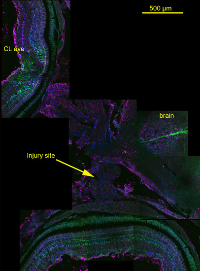
Montage of images showing Tg(gfap:GFP) and cytokeratin localization in the retinas, optic nerves, and a portion of brain obtained from a fish fixed 24 hours post-optic nerve injury. (20x water immersion, NA 0.95). Prominent GFP expression throughout the Müller glia is visible in retinas of eye associated with the injured optic nerve (injury site, arrow) and the contralateral eye (CL eye), and delimiting what appear to be radial glia in a portion of the brain (brain). No GFP expression can be seen in the optic nerve between the retina and optic tract. Cytokeratin (magenta) labeling is apparent in the inner limiting membranes and less intensely in the cytoplasm of cells in the inner plexiform layers of the retinas and in the reticular astrocytes of the optic nerve. Blue label is DAPI indicating nuclei. See Figures 2 and 3 for enlarged views of retina and optic nerve. |