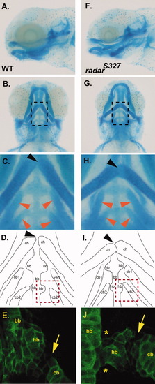
Analysis of pharyngeal arch organization in wild-type and radar mutant larvae. A,B: Lateral and ventral view of Alcian blue stained 5 days postfertilization (dpf) wild-type larvae. C: High magnification of dotted box area in A noting normal articulation of ceratohyals (at joint indicated by black arrow) and normal positions of ceratobranchials (red arrows) D: Camera lucida image outlining the Alcian blue stained cartilages in B. E: Collagen-2α1 staining of the third arch in 5 dpf wild-type larvae to visualize ceratobranchial, basibranchial, and hypobranchial. F,G: Lateral and ventral view of Alcian blue radars327 mutant. H: High magnification of region in panel F demarcated by dotted box showing abnormal articulation of ceratohyals (black arrow) and more sharply angled ceratobranchials (red arrows). I: Camera lucida image of the Alcian blue stained cartilages in E. J: Collagen-2α1 staining of third arch in 5 dpf mutant larvae reveals morphological abnormalities of the hypobranchial (asterix) and hypobranchial/ceratobranchial joint (arrow). ch, ceratohyals; cb, ceratobranchials; bb, basibranchial; hb, hypobranchials.
|
