
FIGURE
Fig. 4
- ID
- ZDB-FIG-100223-38
- Publication
- Hultman et al., 2010 - Differential contribution of direct-developing and stem cell-derived melanocytes to the zebrafish larval pigment pattern
- Other Figures
- All Figure Page
- Back to All Figure Page
Fig. 4
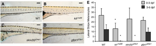
Late-stage melanocyte development in larval melanocyte regeneration mutants. Lateral trunk view at 6 dpf of (A) wild type, (B) kitj1e99, (C) skiv2l2j24e1 and (D) gfpt1j23e1. Scale bars, 100 um. (E) Quantitation of embryonic lateral stripe melanocytes at 3 dpf (light gray) and melanocytes that develop from 3 to 6 dpf (dark gray) in these mutants. Mean value (n = 10 for wild type, kitj1e99 and gfpt1j23e1, n = 4 for skiv2l2j24e1) with error bars representing standard deviation. ∗P-values £ 0.05. |
Expression Data
Expression Detail
Antibody Labeling
Phenotype Data
Phenotype Detail
Acknowledgments
This image is the copyrighted work of the attributed author or publisher, and
ZFIN has permission only to display this image to its users.
Additional permissions should be obtained from the applicable author or publisher of the image.
Reprinted from Developmental Biology, 337(2), Hultman, K.A., and Johnson, S.L., Differential contribution of direct-developing and stem cell-derived melanocytes to the zebrafish larval pigment pattern, 425-431, Copyright (2010) with permission from Elsevier. Full text @ Dev. Biol.