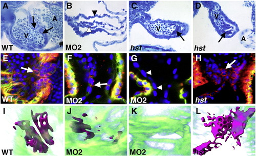
Pdlim7 and Tbx5 are required for valve development. (A–D) 72 hpf sagittal sections stained with methylene blue from wild-type (A), MO2 injected (B), and hst embryos (C, D). Black arrowhead indicates myocardium, white arrowhead indicates endocardium in B. Black arrows depict valve tissue in A, C, D. Head is positioned to the right. (E–H) Ventral views of whole mount embryo immunofluorescence at 72 hpf with zn8 (red, Dm-grasp), Alexa 488 phalloidin (green, actin) and DAPI (blue, nuclei) on wild-type (E), MO2 injected (F, G), and hst (H) embryos. White arrows indicate endocardium with Dm-grasp expression in E, F, H. Arrowheads in G denote endocardial layer lacking Dm-grasp. (I–L) Three-dimensional reconstruction of confocal z-stacks at 72 hpf of wild-type (I; Supplemental movie 2), MO2 injected (J, K; Supplemental movies 3 and 4), and hst (L; Supplemental movie 5) embryos. High power views of endocardial Dm-grasp positive cells at the AV boundary (magenta). Myocardial actin (green) is filtered transparent and DAPI nuclei staining has been removed to enhance Dm-grasp visibility (Amira software, see Materials and methods). V = ventricle. A = atrium. Head is positioned to the top.
|
