
FIGURE
Fig. 5
- ID
- ZDB-FIG-100114-29
- Publication
- Codina et al., 2010 - Loss of Smyhc1 or Hsp90alpha1 function results in different effects on myofibril organization in skeletal muscles of zebrafish embryos
- Other Figures
- All Figure Page
- Back to All Figure Page
Fig. 5
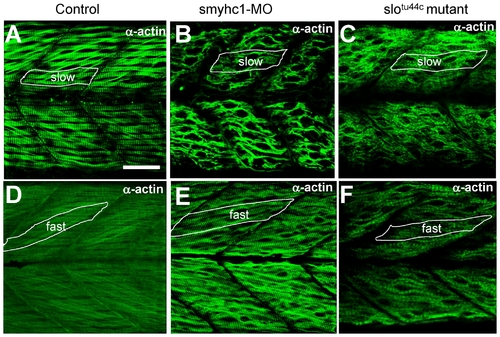
Knockdown of smyhc1 expression or hsp90α1 mutation resulted in defective thin filament organization in skeletal muscles of zebrafish embryos. A–C. Anti-α-actin antibody staining shows the organization of thin filaments in slow muscles of control (A), smyhc1 knockdown (B), or slotu44c mutant (C) embryos at 48 hpf. D, F. Anti-α-actin antibody staining shows the organization of thin filaments in fast muscles of control (D), smyhc1 knockdown (E), or slotu44c mutant (F) embryos at 72 hpf. Scale bar = 25 μm in A. |
Expression Data
| Antibody: | |
|---|---|
| Fish: | |
| Knockdown Reagent: | |
| Anatomical Terms: | |
| Stage Range: | Long-pec to Protruding-mouth |
Expression Detail
Antibody Labeling
Phenotype Data
| Fish: | |
|---|---|
| Knockdown Reagent: | |
| Observed In: | |
| Stage Range: | Long-pec to Protruding-mouth |
Phenotype Detail
Acknowledgments
This image is the copyrighted work of the attributed author or publisher, and
ZFIN has permission only to display this image to its users.
Additional permissions should be obtained from the applicable author or publisher of the image.
Full text @ PLoS One