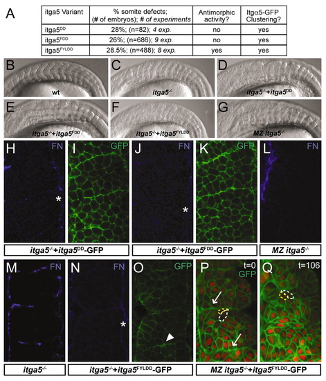
Initial clustering of Itgα5-GFP is independent of FN. (A)In vitro transcribed mRNAs encoding Itgα5-GFP variants were injected at 250 ng/μl into zebrafish embryos derived from Itgα5-/+ parents. Injected embryos were assayed morphologically for somite defects, for antimorphic activity and for Itgα5-GFP clustering. (B-G) Lateral views of the trunk somites in 14- to 18-somite stage embryos. Anterior is left. (B) A wild-type (wt) embryo. (C) An itga5-/-embryo. Somitogenesis in itga5-/- is not rescued by injection of mRNA encoding Itgα5DD-GFP (D), Itgα5FDD-GFP (E) or Itgα5FYLDD-GFP (F). The itga5-/- embryo in C is an uninjected sibling of the embryo in F. Note that the morphological somite defects are enhanced by Itgα5FYLDD expression. (G) Loss of maternal itga5 enhances the zygotic itga5-/- phenotype; however, posterior trunk and tail somites form in these embryos. (H-O) Embryos at the 6- to 8-somite stage. Itgα5DD-GFP does not rescue segmental FN assembly in itga5-/- embryos (H) but clusters along nascent borders (I). Similarly, Itgα5FDD-GFP does not rescue segmental FN assembly in itga5-/- embryos (J) but clusters along nascent borders (K). (L) MZ itga5-/- embryos lack segmental FN. FN localization in itga5-/- (M) and an itga5-/- embryo injected with Itgα5FYLDD (N). (O) Itgα5FYLDD-GFP localization in the embryo shown in N. Note that the FN-matrix defects are enhanced by injection of Itgα5FYLDD, with matrix only forming along the surface of the paraxial mesoderm (asterisks). Nonetheless, Itgα5FYLDD-GFP clustering is observed (arrowhead). (P,Q) Two time points (indicated in minutes) of Itgα5FYLDD-GFP localization in an 8- to 10-somite stage embryo lacking maternal and zygotic itga5 (MZ itga5-/-). A functional ligand-binding domain is not necessary for initial clustering (arrows) but is required to maintain clustering. For reference, the same two cells are outlined in yellow and white. (H-Q) Anterior is up.
|
