|
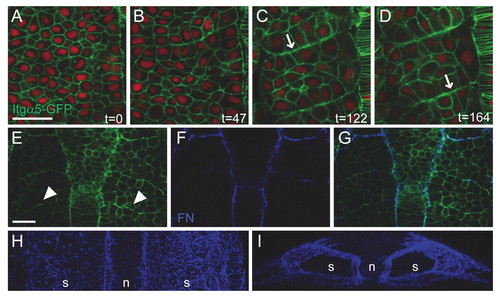
Itgα5-GFP clustering and FN matrix assembly during somitogenesis in zebrafish. (A-D) Four time points (indicated in minutes) showing Itgα5-GFP localization during somite formation. Itgα5-GFP (green) is distributed along the cortex in mesenchymal presomitic cells (A) but clusters to the basal side of columnar border cells (B-D, arrows). Nuclei are red. (E-G)Itgα5-GFP (E), FN (F) and overlay (G). Nascent borders show Itgα5-GFP clustering (arrowheads in E) but no FN immunostaining. (H,I) Three-dimensional reconstruction showing FN matrix along the surface of the paraxial mesoderm. (A-H) Dorsal views, anterior is up. (I) Rotation of H showing a transverse view of the presomitic mesoderm, dorsal is up. Embryos are at the 8- to 10-somite (A-D,H,I) or 5- to 6-somite (E-G) stage. n, notochord; s, somites. Scale bars: 30 μm.
|