
Fig. 7
- ID
- ZDB-FIG-090508-15
- Publication
- Riley et al., 1997 - A critical period of ear development controlled by distinct populations of ciliated cells in the zebrafish
- Other Figures
- All Figure Page
- Back to All Figure Page
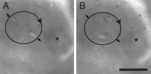
Non-Brownian motion of otolith precursor particles. (A and B) Phase-contrast images captured on time-lapse video show two particles moving in a vortex (large circular arrow) just anterior and medial to the posterior otolith. Although the otolith is out of focus, its position can be discerned to the right of the vortex (asterisk). (A) Small arrows mark the initial positions of two particles orbiting on opposite sides of the vortex. (B) Approximately 0.1 s later, the particles have moved several micrometers in a clockwise orbit. The particles completed 10 – 12 rotations before being ejected from the vortex. Observation of this and other vortices revealed that small particles typically rotate faster than large particles, and that the overall speed of rotation fluctuates over time (not shown). Images were captured from a videotape of the same wild-type ear shown in Figs. 5B – 5F. Anterior is to the left and dorsal is upward. Scale bar, 5 μm. |
Reprinted from Developmental Biology, 191(2), Riley, B.B., Zhu, C., Janetopoulos, C., and Aufderheide, K.J., A critical period of ear development controlled by distinct populations of ciliated cells in the zebrafish, 191-201, Copyright (1997) with permission from Elsevier. Full text @ Dev. Biol.