
Fig. 6
- ID
- ZDB-FIG-090508-14
- Publication
- Riley et al., 1997 - A critical period of ear development controlled by distinct populations of ciliated cells in the zebrafish
- Other Figures
- All Figure Page
- Back to All Figure Page
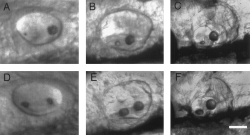
Otolith development following modification. (A–C) Development of the same wild-type ear depicted in Figs. 5B–5F following enhancement of the posterior otolith with laser tweezers. (A) At 24 h, approximately 2 h after laser treatment, the posterior otolith is still many times the size of the anterior otolith. At 2 (B) and 6 days (C), the posterior otolith is nearly normal in size, but the anterior otolith remains much smaller than normal. (D–F) Development of a wild-type control embryo at 24 h (D), 2 days (E), and 6 days (F). Anterior is to the left and dorsal is upward in all panels. Scale bar, 25 (A and D), 45 (B and E), and 100 μm (C and F). |
Reprinted from Developmental Biology, 191(2), Riley, B.B., Zhu, C., Janetopoulos, C., and Aufderheide, K.J., A critical period of ear development controlled by distinct populations of ciliated cells in the zebrafish, 191-201, Copyright (1997) with permission from Elsevier. Full text @ Dev. Biol.