
Fig. 6
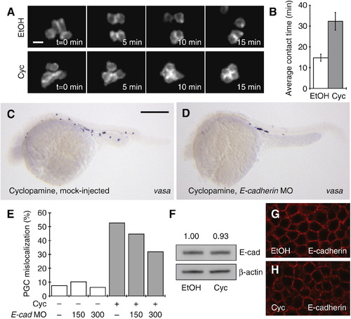
Cyclopamine perturbs PGC migration in part through dysregulated cell adhesion. (A) Time-lapse imaging of PGC interactions in ethanol- or cyclopamine-treated Tg(kop:EGFP-F-nanos1-3′UTR) embryos. (B) Average duration of each PGC–PGC contact for both treatment conditions. Data are the averages of 35 ethanol-treated and 45 cyclopamine-treated PGCs, and error bars represent the standard error of the mean. (C, D) Distribution of vasa-positive PGCs in embryos injected with either water or an E-cadherin MO (300 pg/embryo) at the one-cell stage and then treated with 50 μM cyclopamine until 24 hpf. Scale bar: 200 μm. (E) Quantification of PGC mislocalization in embryos injected with varying doses of the E-cadherin MO and treated with cyclopamine or an ethanol vehicle control. At least 500 PGCs were scored for each experimental condition. (F) Western analysis of E-cadherin expression in embryos treated with ethanol or cyclopamine from the one-cell stage to the sphere stage (6 hpf). Quantification of E-cadherin expression normalized to a β-actin loading control is shown. (G, H) Confocal immunofluorescence imaging of E-cadherin expression and localization in identically treated embryos. Scale bars: A, 20 μm; C, D, 200 μm. |
Reprinted from Developmental Biology, 328(2), Mich, J.K., Blaser, H., Thomas, N.A., Firestone, A.J., Yelon, D., Raz, E., and Chen, J.K., Germ cell migration in zebrafish is cyclopamine-sensitive but Smoothened-independent, 342-354, Copyright (2009) with permission from Elsevier. Full text @ Dev. Biol.