
Fig. 4
- ID
- ZDB-FIG-090401-4
- Publication
- Sonawane et al., 2009 - Lgl2 and E-cadherin act antagonistically to regulate hemidesmosome formation during epidermal development in zebrafish
- Other Figures
- All Figure Page
- Back to All Figure Page
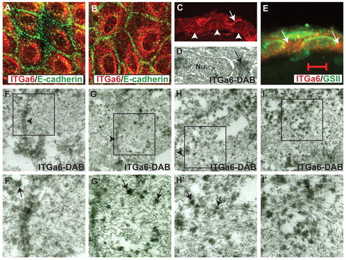
Lgl2 mediates targeting of Itga6 to the plasma membrane during hemidesmosome formation in basal epidermal cells. (A-C,E) Immunostaining of lgl2 mutant epidermis with anti-Itga6 (red) and anti-E-cadherin (green) antibodies (A,B), anti-Itga6 antibody (C), or anti-Itga6 antibody (red) and lectin GS II (green) in section (E). In mutant larvae at 4-5 dpf, Itga6 exhibits diffuse cytoplasmic staining along with strong perinuclear accumulation (A,B). This perinuclear Itga6 staining is not due to Itga6 accumulation in the Golgi apparatus (E). Basal and lateral Itga6 localisation is lost in lgl2 larvae (C); arrowheads mark the basal domain and the arrow marks the apical domain juxtaposed to a peridermal cell where the Itga6 staining occasionally persists. (D,F-I′) Immunoelectron microscopy using anti-Itga6 antibody and nickel-enhanced DAB in wild-type (F,F′) and lgl2 mutant (D,G-I′) larvae. F′-I′ are 2x enlargements of the boxed regions in F-I. The perinuclear accumulation of Itga6 is evident in immunoelectron micrographs (arrow in D). In wild-type larval epidermis at 4 dpf, electron-dense Itga6 vesicles are relatively rare (F) around the lateral domain. By contrast, in lgl2 mutant larvae at 4 dpf, Itga6 vesicles are frequently present in the lateral cortex (G,H) and in the cytoplasm in general (I). Arrowheads in F-I indicate the lateral domain. In F′-I′, some of the Itga6 vesicles are indicated by arrows. Scale bar: 13.5 μm in A,B; 27 μm in C; 5 μm in E; 200 nm in F-I. |