
Fig. 4
- ID
- ZDB-FIG-090331-6
- Publication
- Lin et al., 2009 - Differential expression of zebrafish gpia and gpib during development
- Other Figures
- All Figure Page
- Back to All Figure Page
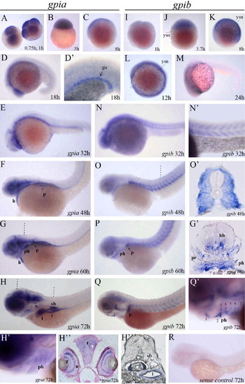
Differential expression patterns of gpia and gpib during zebrafish embryonic development. Whole-mount in situ hybridized embryos showing gpia (A–H), gpib (I–Q) mRNA expression, and a control embryo hybridized with gpia sense probe (R). The hour post fertilization for each embryo is shown at the bottom corner. (D′, H′, N′, Q′) are higher magnification views of (D, H, N, Q) respectively. (G′, H″, H′″, O′) are transverse cryosections through the embryos in (G, H, O) at the levels indicated by dotted lines, respectively. Section in (H″) was counterstained with Nuclear Fast Red. Abbreviations: gu, primitive gut; h, heart; p, pectoral fin; ph, pharyngeal arches; sb, smooth muscle cell layer of developing swim bladder; l, liver; i, intestinal epithelia; t, tectum; g, ganglion cell layer; n, inner nuclear layer of the retina; ysn, yolk syncytial nuclei; 1–7 ph, the first to the seventh pharyngeal arch. |
| Genes: | |
|---|---|
| Fish: | |
| Anatomical Terms: | |
| Stage Range: | 4-cell to Protruding-mouth |
Reprinted from Gene expression patterns : GEP, 9(4), Lin, W.W., Chen, L.H., Chen, M.C., and Kao, H.W., Differential expression of zebrafish gpia and gpib during development, 238-245, Copyright (2009) with permission from Elsevier. Full text @ Gene Expr. Patterns