|
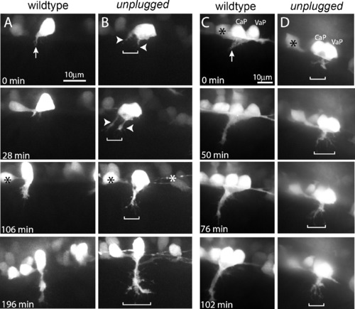
unplugged Restricts Navigating Growth Cones to a Central Muscle Zone (A–D) Still images from time-lapse movies showing the initial migration of single CaP axons (A and B) or CaP/VaP pair axons (C and D) from the spinal cord into the myotome. Arrows point to the single wild-type CaP growth cone (A) and to the tightly fasciculated wild-type CaP/VaP growth cones (C). In contrast, unplugged CaP neurons form extensive filopodia and even multiple growth cones (arrowheads) that occupy a broader area (brackets, [B]). Similary, mutant CaP/VaP growth cones appear defasciculated and occupy a broader area compared to wild-type. Asterisks indicate interneurons also labeled by the Tg(Hb9:GFP) transgene.
|