
Fig. 1
- ID
- ZDB-FIG-090320-36
- Publication
- Sims Jr et al., 2009 - Connexin43 regulates joint location in zebrafish fins
- Other Figures
- All Figure Page
- Back to All Figure Page
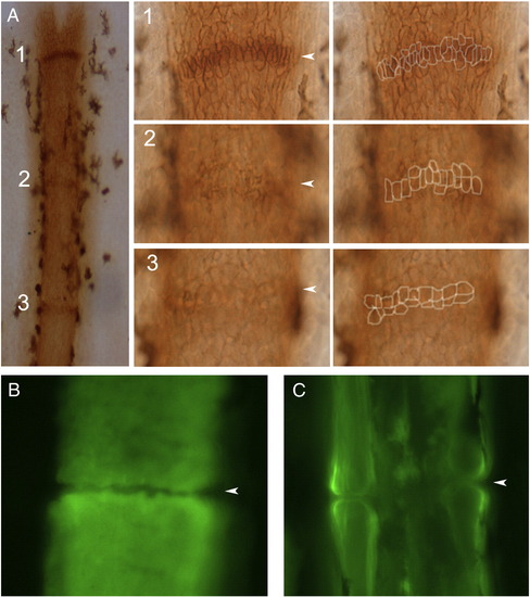
Joint formation in zebrafish fin rays. (A) Fins were stained with the osteoblast marker ZNS5, and detected using HRP (brown). At this level joints are detected as condensations of ZNS5-positive cells, which appear with differing maturities along the proximal–distal axis. Joint 1 is the most-distal joint and the least mature. At higher magnification distinct morphologies of ZNS5-positive cells can be observed (and outlines of the ZNS5-positive cells are shown to the right). (B) The morphology of the bone matrix in the newest joint is observed using calcein. (C) The morphology of the bone matrix in a fully mature joint is observed using calcein. Arrowheads point to joints. |
| Antibody: | |
|---|---|
| Fish: | |
| Anatomical Term: | |
| Stage: | Adult |
Reprinted from Developmental Biology, 327(2), Sims Jr, K., Eble, D.M., and Iovine, M.K., Connexin43 regulates joint location in zebrafish fins, 410-418, Copyright (2009) with permission from Elsevier. Full text @ Dev. Biol.