|
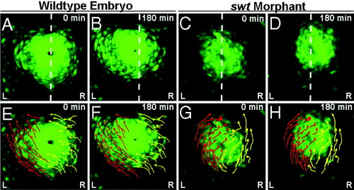
Asymmetric atrial cell migration promotes rotation of the cardiac cone. (A–H) Dorsal views of the heart in Tg(cmlc2:egfp) embryos between 18–21 hpf. (A–D) Dotted white lines indicate the embryonic midline. (E–H) Arrows indicate trajectories of left (red) and right (yellow) cells. A, E, C, and G are the first frames of time lapses (0 min), and B, F, D, and H are the final frames (180 min). A, B, E, and F are frames from a time lapse of a single WT embryo, and C, D, G, and H are frames from a time lapse of a single swt morphant. In WT embryos, the left atrial myocardium migrates asymmetrically along the left of the cone toward the left and anterior (E and F). The right cardiac cells also migrate toward the anterior and left, but rather than sweeping along the lateral edge of the cone, these cells migrate toward the lumen (E and F). In swt morphants the L/R directionality of these cellular trajectories is reversed (C, D, G, and H). L, left; R, right.
|