
Fig. 1
- ID
- ZDB-FIG-090220-29
- Publication
- Baxendale et al., 2009 - Expression screening and annotation of a zebrafish myoblast cDNA library
- Other Figures
- All Figure Page
- Back to All Figure Page
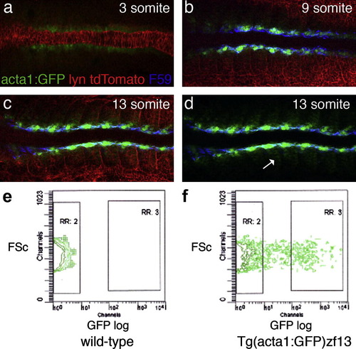
GFP expression and FACS analysis of Tg(acta1:GFP)zf13 embryos. (a–d) Dorsal view of flat-mounted Tg(acta1:GFP)zf13 embryos at 3, 9 and 13 somite stages, anterior to the left. Embryos were injected with Lyn:tdTomato RNA to highlight the cell membranes (red) and immunohistochemistry with F59, specific for slow myosin heavy chain (Devoto et al., 1996), labeled the slow myoblast cells blue. (a) Three somite stage embryo. GFP is weakly expressed in the adaxial cells flanking the notochord. No F59 stain can be detected at this stage. (b) Nine somite stage embryo. GFP is seen in adaxial cells, which are co-stained with F59. (c) Thirteen somite stage embryo. The strongest GFP expression is medial next to the notochord. The most anterior adaxial cells are elongated and have slow twitch myofibrils detected by F59. Weaker GFP expression is seen in the fast twitch fibre precursors. (d) The same picture as (c) with the red channel omitted. The weaker GFP-expressing cells are seen clearly in the posterior half of the somite; the site of early myoD expression where the first fast myoblast cells differentiate. (e and f) FACS analysis of dissociated Tg(acta1:GFP)zf13 embryos. Each plot shows the forward scatter (FSc) plot of the single cell population (x-axis) against GFP fluorescence (y-axis). In wild-type embryos (1E) no fluorescence is detected. In GFP-expressing embryos cells show a range of fluorescence with 12.5% of cells expressing GFP strongly (RR:3). |
| Gene: | |
|---|---|
| Antibody: | |
| Fish: | |
| Anatomical Terms: | |
| Stage Range: | 1-4 somites to 10-13 somites |
Reprinted from Gene expression patterns : GEP, 9(2), Baxendale, S., Chen, C.K., Tang, H., Davison, C., Hateren, L.V., Croning, M.D., Humphray, S.J., Hubbard, S.J., and Ingham, P.W., Expression screening and annotation of a zebrafish myoblast cDNA library, 73-82, Copyright (2009) with permission from Elsevier. Full text @ Gene Expr. Patterns