
Fig. 5
- ID
- ZDB-FIG-090113-81
- Publication
- Suhr et al., 2009 - Highly-restricted, cell-specific expression of the simian CMV-IE promoter in transgenic zebrafish with age and after heat shock
- Other Figures
- All Figure Page
- Back to All Figure Page
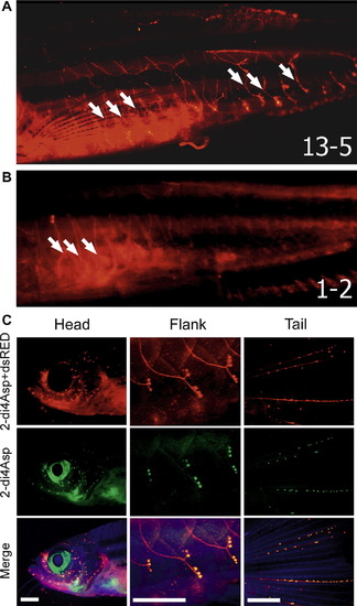
(A) Fluorescent image of the flank of a sCMV:dsRed line 13-5 fish at 5 months showing expression in the anterior and posterior lateral line nerves (white arrows). (B) Flank of a sCMV:dsRed line 1-2 fish age matched to the fish in (A) to show variation in the intensity of expression in the lateral line and appendicular muscle. (C) The lateral line system in a 2-month old sCMV:dsRed line 13-5 fish in three areas: head, trunk (flank), and tail. Lateral line fibers are shown with dsRed expression (red), the lateral line neuromast dye DASPEI (red/green in neuromasts only), and autofluorescence (blue – included to facilitate orientation). DASPEI stained hair cells of the neuromasts appear as bright dots in the green channel. Faint green fluorescence in the fibers proximal to neuromasts in the 2Di4Asp panels is due to low levels of DASPEI back-labeling. Scale bars = 1 mm (anterior left in all images). |
| Gene: | |
|---|---|
| Fish: | |
| Anatomical Terms: | |
| Stage Range: | Days 45-89 to Adult |
Reprinted from Gene expression patterns : GEP, 9(1), Suhr, S.T., Ramachandran, R., Fuller, C.L., Veldman, M.B., Byrd, C.A., and Goldman, D., Highly-restricted, cell-specific expression of the simian CMV-IE promoter in transgenic zebrafish with age and after heat shock, 54-64, Copyright (2009) with permission from Elsevier. Full text @ Gene Expr. Patterns