
Fig. 3
- ID
- ZDB-FIG-090113-79
- Publication
- Suhr et al., 2009 - Highly-restricted, cell-specific expression of the simian CMV-IE promoter in transgenic zebrafish with age and after heat shock
- Other Figures
- All Figure Page
- Back to All Figure Page
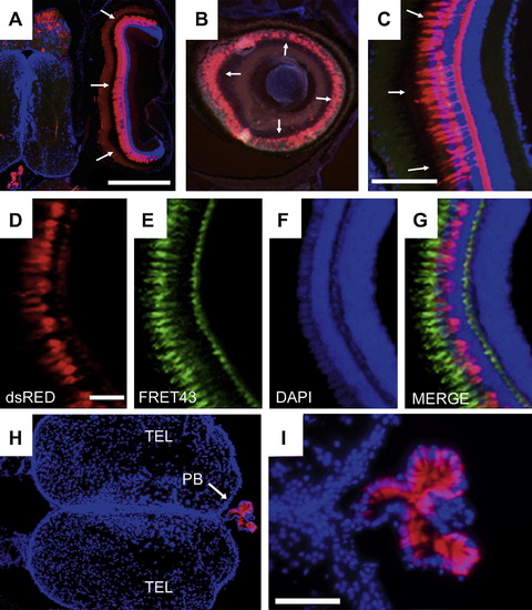
(A) Horizontal section through the head of an adult transgenic fish showing the brain and adjacent eye with strong dsRed-expressing cells apparent in the retina (arrows). Blue is DAPI stain and anterior is up. (B) Fluorescent cells appear as a ring (indicated by arrows) in para-sagittal sections of retina. (C) Cross-section of DAPI-stained retina revealing that fluorescent cells have the position and morphology of photoreceptors. (D–G) Cellular analysis of reporter-expressing cells in the adult fish retina showing dsRed fluorescence (D), FRET43 immunolabeling (E), DAPI (F), and a merge of all three channels in (G). (H and I) Low-(H) and high-(I) magnification images of dsRed-positive cells (red) and DAPI staining (blue) in horizontal sections of an adult transgenic fish brain showing the pineal body. In (H and I), anterior is to the left. Scale bars: A/B = 500 μm; C = 125 μm; D–G = 50 μm; H = 250 μm; I = 100 μm. All images are of line sCMV:dsRed 2-1. |
| Gene: | |
|---|---|
| Antibody: | |
| Fish: | |
| Anatomical Terms: | |
| Stage: | Adult |
Reprinted from Gene expression patterns : GEP, 9(1), Suhr, S.T., Ramachandran, R., Fuller, C.L., Veldman, M.B., Byrd, C.A., and Goldman, D., Highly-restricted, cell-specific expression of the simian CMV-IE promoter in transgenic zebrafish with age and after heat shock, 54-64, Copyright (2009) with permission from Elsevier. Full text @ Gene Expr. Patterns