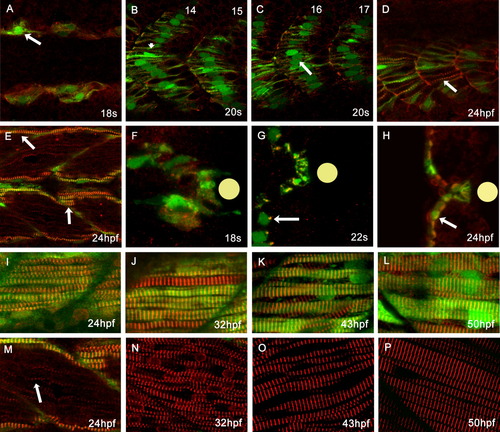
Normal myofibrillogenesis in Tg(smyhc1:GFP)i104 transgenic embryos. A: Dorsal view of an 18 somite stage embryo showing the first appearance of striated α-Actinin (red: arrow) in slow muscle fibres (marked by GFP expression). B,C: Lateral aspect of a 20-hpf embryo: α-Actinin was organised as a clear z-band (arrowhead) in anterior somites (B), but not in more posterior somites (C, arrow). D,E: Lateral aspect of 24-hpf embryo: posterior slow fibres have clearly discernible striated thin myofibrils (D) while in more anterior somites the myofibrils have become quite thick (E, arrows). F,G: Transverse sections at successive stages reveal that in slow fibre progenitors, prior to their migration away from the notochord (indicated by the yellow disc) α-Actinin distribution is quite diffuse (F), but as migration proceeds, it becomes localised to one side of the nucleus (G, arrow) and 24 hpf reveals the fully assembled myofibrils (H). I-P: Pairs of images of lateral aspects of the surface slow muscle and the underlying fast muscle at successively later stages of development: fast muscle myotubes started to express and localise α-Actinin as a single file of myofibrils at 24 hpf, much later than in slow muscle fibres (M: arrow). Myofibrillogenesis proceeds rapidly in both slow and fast lineages between 32-50 hpf.
|
