|
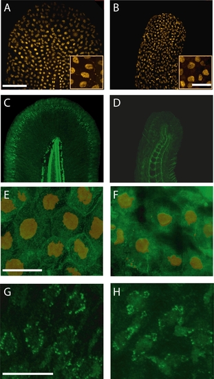
Ap1s1 knockdown is associated with abnormal distribution of laminin and cadherin. Despite changes in the size and shape of the tail, p63-labelled nuclei (orange) of basal keratinocytes were present in KD larvae. (B). The insets show enlarged views of small groups of keratinocytes. Immunolabelling for laminin (green) in WT (C) showed normal distribution of the basement membrane along the fin fold margin, whereas in the KD larvae the residual laminin labeling was diffuse and disorganized (D). Compared to WT keratinocytes (E) labeled with p63 (orange), cadherin (green) is decreased at the cell membrane of the KD larvae (F). However, the localization of cytokeratin in WT (G) and in KD larvae (H) appeared similar. Scale bars: 10 μm, Insets in A, B, = 20 μm.
|