|
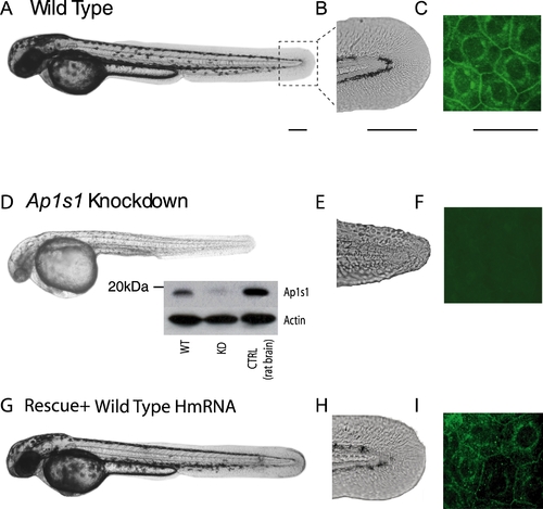
Morphological phenotype of Ap1s1 knockdown zebrafish is rescued by over expression of human AP1S1. Transmitted light images of 48 hpf Ap1s1 KD larvae show their smaller size, reduced pigmentation (D) and skin disorganization (E) compared to the WT (A, B) and the rescued larvae (G,H). Immunofluorescence of wholemount zebrafish using anti-Ap1s1 antibody showing localization of Ap1s1 to the plasma membrane (C, polygonal) and to a well defined perinuclear ring, in both normal (C) and rescued larvae (G), whereas only a residual and diffuse staining could be observed in KD larvae (F). Western blot analysis (D, inset) indicates nearly complete knockdown of Ap1s1 protein (WT = wild-type, KD = knockdown, CTRL = control rat brain proteins). To normalize the western blot analysis, proteins extracted from WT, KD and CTRL larvae were incubated with anti-actin. Scale bars in (A, D, G) = 100 μm, (B, C, E, F, H, I-Ciii) = 50 μm.
|