
FIGURE
Fig. 7
- ID
- ZDB-FIG-081222-18
- Publication
- Kagemann et al., 2008 - Repeated, noninvasive, high resolution spectral domain optical coherence tomography imaging of zebrafish embryos
- Other Figures
- All Figure Page
- Back to All Figure Page
Fig. 7
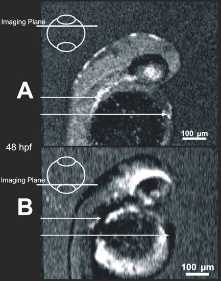
Blood within vessels creates shadow artifacts in C-mode slices below the vessels. Images of the same 48 hpf embryo obtained in a shallow slab location (A) and a deep location (B) allow visualization of blood, which is highly scattering (A, bright locations, arrows), and the resultant shadows mask structure in deeper slabs (B, dark locations, arrows). |
Expression Data
Expression Detail
Antibody Labeling
Phenotype Data
Phenotype Detail
Acknowledgments
This image is the copyrighted work of the attributed author or publisher, and
ZFIN has permission only to display this image to its users.
Additional permissions should be obtained from the applicable author or publisher of the image.