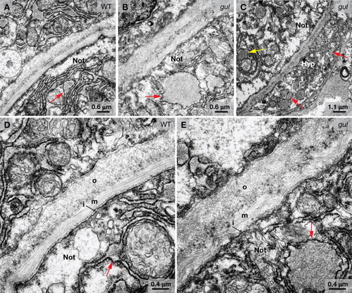
Electron microscopy reveals notochord abnormalities in gulm208 mutants. A-E: Transmission electron micrographs of truncal cross-sections from embryos at 24 hpf. A: Notochord sheath and vacuolated notochord cell of a wild-type embryo, with small areas of protein accumulation visible in the rough endoplasmic reticulum (arrow). B: Notochord sheath and vacuolated notochord cell of a gulm208 mutant with large circular aggregates of protein in the rough endoplasmic reticulum (arrow). C: Notochord and hypochord of a gulm208 mutant with large circular aggregrates of protein in the hypochord (red arrows) and notochord (yellow arrow). D: Notochord sheath of a wild-type embryo with inner (i), medial (m), and outer (o) layers indicated. Small areas of protein accumulation are visible in the rough endoplasmic reticulum (red arrow). E: Notochord sheath of a gulm208 mutant where the collagen fibrils in the medial (m) layer appear disorganized. A large circular aggregate of protein is visible in the rough endoplasmic reticulum (red arrow). Not, vacuolated notochord cell; Hyp, hypochord.
|
