FIGURE

Fig. 1

ID
ZDB-FIG-081205-18
Publication
Snelson et al., 2008 - Formation of the asymmetric pineal complex in zebrafish requires two independently acting transcription factors
Other Figures
All Figure Page
Back to All Figure Page
Fig. 1

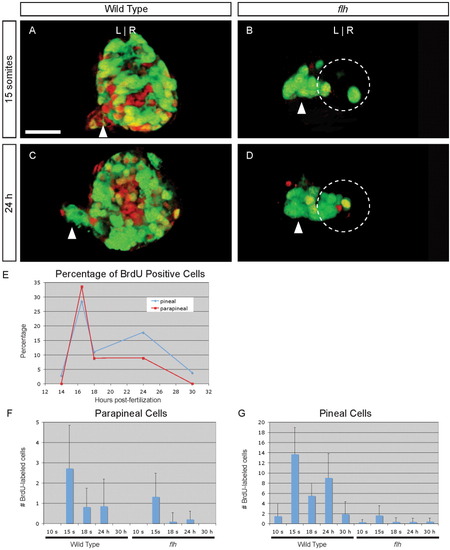
Cell division of parapineal precursors is largely complete by 18 s. A-D: Dorsal views of 4 d larvae, with BrdU (red) and foxd3:gfp (green) labeling in the epithalamus, following a BrdU pulse at (A, B) 15 s or (C, D) 24 hr stage in (A, C) WT or (B, D) flh mutant embryos. Dashed circles in B, D indicate the pineal cells. Scale bar = 25 μM. E: Line graph of the percentage of WT parapineal (red line) or pineal (blue line) cells that are labeled at 4 d, following a BrdU pulse at the indicated developmental stages. F: Bar graph showing the absolute number of BrdU-positive parapineal cells in WT or flh embryos. G: BrdU-positive pineal cells in WT or flh embryos.

Expression Data
Gene:
Antibody:
Fish:
Anatomical Term:
Stage Range: 14-19 somites to Prim-5

Expression Detail
Antibody Labeling
Phenotype Data

Phenotype Detail
Acknowledgments
This image is the copyrighted work of the attributed author or publisher, and ZFIN has permission only to display this image to its users. Additional permissions should be obtained from the applicable author or publisher of the image. Full text @ Dev. Dyn.