
Fig. 1
- ID
- ZDB-IMAGE-081205-15
- Genes
- Antibodies
- Publication
- Snelson et al., 2008 - Formation of the asymmetric pineal complex in zebrafish requires two independently acting transcription factors
- All Figures
- Figures for Snelson et al., 2008
|
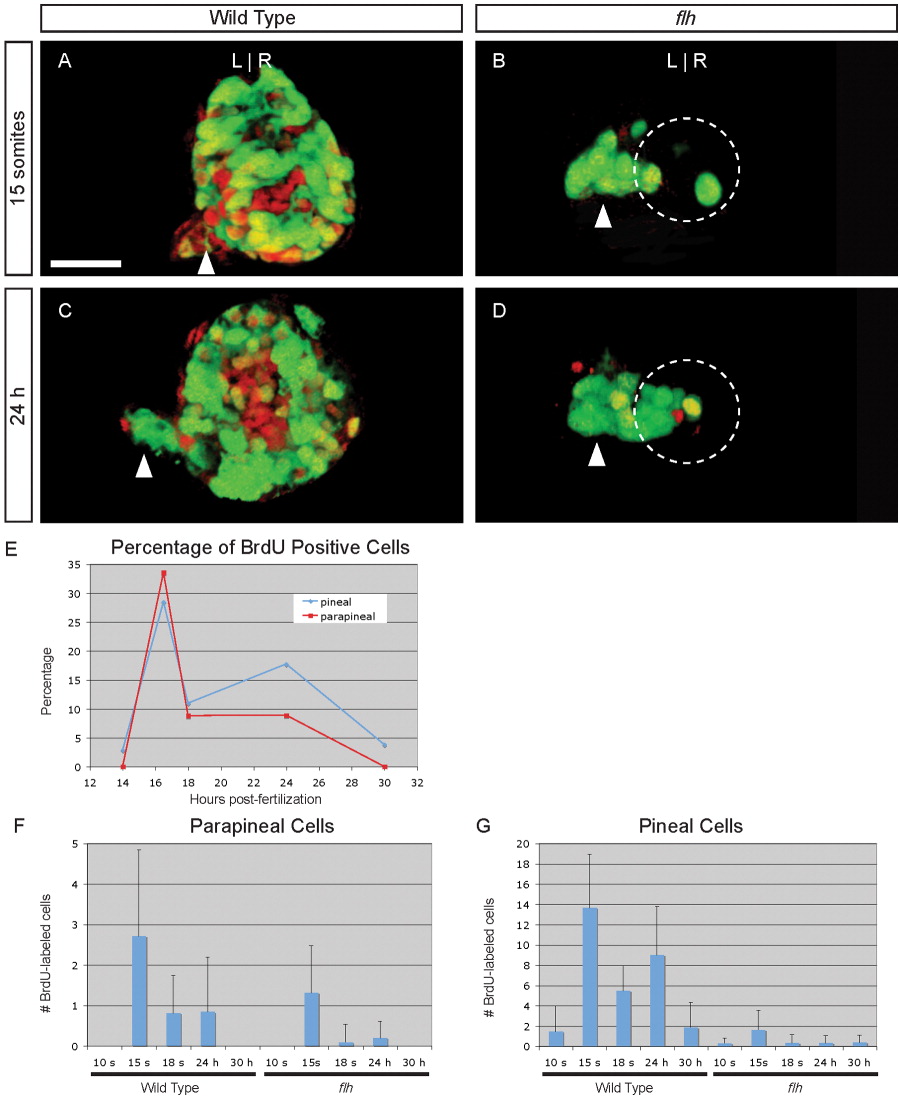
Fig. 1 Cell division of parapineal precursors is largely complete by 18 s. A-D: Dorsal views of 4 d larvae, with BrdU (red) and foxd3:gfp (green) labeling in the epithalamus, following a BrdU pulse at (A, B) 15 s or (C, D) 24 hr stage in (A, C) WT or (B, D) flh mutant embryos. Dashed circles in B, D indicate the pineal cells. Scale bar = 25 μM. E: Line graph of the percentage of WT parapineal (red line) or pineal (blue line) cells that are labeled at 4 d, following a BrdU pulse at the indicated developmental stages. F: Bar graph showing the absolute number of BrdU-positive parapineal cells in WT or flh embryos. G: BrdU-positive pineal cells in WT or flh embryos.