
Fig. 3
- ID
- ZDB-FIG-081105-17
- Publication
- Lewis et al., 1999 - Control of muscle cell-type specification in the zebrafish embryo by hedgehog signalling
- Other Figures
- All Figure Page
- Back to All Figure Page
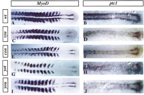
ptc1 and MyoD expression at 10–15 somites. (A) Expression of myoD in 15-somite and (B) ptc1 in 10- to 12-somite stage wild-type (wt) embryos. At the 12- to 15-somite stage, the expression of myoD is reduced in the adaxial cells of embryos homozygous for syu (C), con (E), yot (G), and you (I). Note the persistent expression in the tailbud in yot homozygotes (G). Lateral somite expression of myoD is also slightly reduced in the most rostral somites, most notably at this stage in syu, con, and yot. Expression of ptc1 is also reduced in the adaxial cells of syu (D), con (F), yot (H), and you (J) homozygotes at the 10- to 12-somite stage. |
| Genes: | |
|---|---|
| Fish: | |
| Anatomical Terms: | |
| Stage Range: | 10-13 somites to 14-19 somites |
Reprinted from Developmental Biology, 216(2), Lewis, K.E., Currie, P.D., Roy, S., Schauerte, H., Haffter, P., and Ingham, P.W., Control of muscle cell-type specification in the zebrafish embryo by hedgehog signalling, 469-480, Copyright (1999) with permission from Elsevier. Full text @ Dev. Biol.