Fig. 5
- ID
- ZDB-FIG-081028-5
- Publication
- Prykhozhij et al., 2008 - Distinct roles of Shh and Fgf signaling in regulating cell proliferation during zebrafish pectoral fin development
- Other Figures
- All Figure Page
- Back to All Figure Page
|
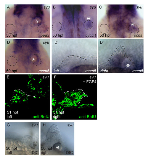
Implantation of FGF4-soaked beads rescues G1- and S-phase cell-cycle gene expression and S-phase progression in sonic-you fin buds. Fin buds on the right hand side of sonic-you embryos were implanted with FGF4-soaked heparin beads at 29–32 hpf, grown until 50 hpf and fixed (A-D, F-H). For anti-BrdU staining, embryos were first implanted and then injected with 10 mM BrdU solution at 38 hpf before fixation at 50 hpf. Sonic-you embryos show upregulation of pea3 (A), cyclinD1 (B), pcna (C) and mcm5 (D) expression in response to the FGF4-soaked beads on the implanted side. Fin buds are outlined by dotted lines in panels A to D. A non-implanted fin bud on the left hand side shows no mcm5 expression (D′), while an implanted fin bud on the right hand side of the same embryo (D″) shows restored mcm5 expression. A non-implanted fin bud shows few BrdU-labeled nuclei (E), while an FGF4 bead-implanted fin bud (F) has extensive BrdU labeling (sections of both sides of 10 bead-implanted embryos were analysed). Fin buds implanted with FGF4 beads show increased outgrowth (D″, F, H), compared to non-implanted control fin buds (D′, E, G). |

