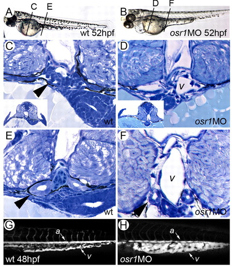
Loss of pronephric epithelial differentiation and vascular expansion in osr1 morphants. Control (A,C,E,G) and osr1 morphants (B,D,F,H) at 52 hpf. (A) Wild-type 52 hpf embryo showing position of histological sections in C and E. (B) osr1 morphant embryo showing position of histological sections in D and F. (C) Cross-section at the level of the fin buds (inset) shows normal glomerular structure (arrowhead) and connecting pronephric tubules. (D) Cross-section at the level of the fin buds in an osr1 morphant (inset) shows absence of pronephric tubules and glomerulus and expansion of cardinal vein (v). (E) In more posterior sections of wild-type embryos, the pronephric epithelial tubules (arrowhead) and cardinal vein are of roughly similar dimensions. (F) In osr1 morphants, pronephric tubules are reduced in diameter compared with wild-type (arrowhead) and the cardinal vein (v) is significantly expanded. (G) Angiogram of wild-type embryo trunk and tail region highlights the aorta (a) and the common tail vein (v). (H) Angiogram of an osr1 morphant highlights a grossly expanded venous plexus in the tail (v). Intersomitic vessels were present in osr1 morphants but are not shown in the confocal sections used in this projection.
|
