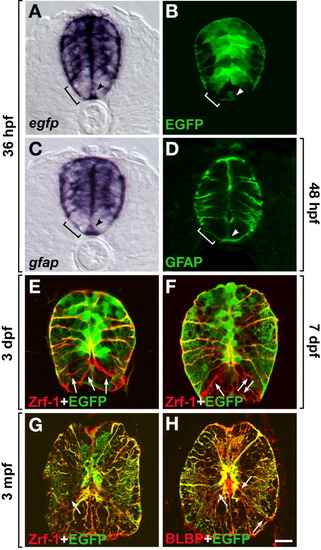
gfap+ cells are radial glia in embryonic and postembryonic zebrafish spinal cord. All images are transverse sections of spinal cords, dorsal to top. A,B: Spinal cords of 36 hours postfertilization (hpf) Tg(gfap:GFP) embryos labeled by gfap (A) and egfp (B) RNA in situ hybridization. C: gfap RNA expression at 36 hpf. D: Glial fibrillary acidic protein (GFAP) expression detected by immunocytochemistry at 48 hpf. Brackets in A-D mark ventral spinal cord cells that do not express the endogenous gfap gene or the reporter transgene. Arrowheads mark floor plate. E-G: Anti-Zrf-1 antibody labeling (red) and enhanced green fluorescent protein (EGFP) fluorescence (green) of Tg(gfap:GFP) animals at 3 days postfertilization (dpf), 7 dpf, and 3 months. Numerous Zrf-1+ EGFP+ radial glia appear as yellow fibers. Arrows indicate Zrf-1+ GFAP- radial glial cells in ventral most spinal cord. H: Anti-brain-lipid-binding protein (BLBP) antibody (red) labeling of 3-month-old adult Tg(gfap:GFP) fish. BLBP+ EGFP+ radial glia appear yellow. Arrows indicate BLBP+ EGFP- radial fibers in ventral spinal cord. Scale bar = 20 μM in A-F, 80 μM in G,H.
|
