
FIGURE
Fig. 4
- ID
- ZDB-FIG-080730-5
- Publication
- Cao et al., 2008 - Hypoxia-induced retinal angiogenesis in zebrafish as a model to study retinopathy
- Other Figures
- All Figure Page
- Back to All Figure Page
Fig. 4
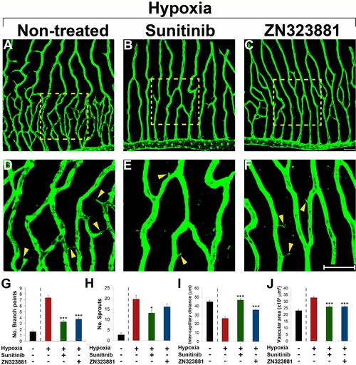
Inhibition of retinal neovascularization by orally active anti-VEGF drugs. Adult fli-EGFP-Tg zebrafish were exposed to 10 % hypoxia in the absence (A) or presence of sunitinib (B and E) or ZN323881 (C and F) anti-VEGF small molecules for 14 days. Retinal neovascularization was analyzed using whole-mount confocal analysis and quantified as branching points (G), numbers of sprouts (H), intercapillary distances (I), and total vascularization area (J). Yellow arrowheads point to vascular sprouts. Data represents mean determinants of 11–29 randomized samples. *p<0.05. ***p<0.001. Bar = 50 μm. |
Expression Data
Expression Detail
Antibody Labeling
Phenotype Data
Phenotype Detail
Acknowledgments
This image is the copyrighted work of the attributed author or publisher, and
ZFIN has permission only to display this image to its users.
Additional permissions should be obtained from the applicable author or publisher of the image.
Full text @ PLoS One